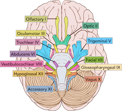
The Cranial Nerves - A Brief Walkthrough
The Cranial Nerves are twelve pairs of nerves originating in the brain (as given by the name "cranial" - showing that they are located inside the cranium.
These nerves are of extreme importance due to the plethora of functions associated with them (ranging all the way from smell to balance) and the wide array of effects it has on our body.
One of each pair are located on each side of the brain, and are numbered in roman numerals I through XII. These are often labeled as CN I, CN II, and so on. The first two cranial nerves, the olfactory nerve and the optic nerve, are located in the cerebrum (located in the front/anterior area of the skull) , and the rest in the brainstem (posterior part of the brain which is continuous with the spinal cord)
CN I --> OLFACTORY NERVE: To do with the transmission of sensory information regarding smells. Smells are analysed in the olfactory bulb which stimulates the nerve cells present there to pass impulses to the olfactory tract and finally to the the brain where they are recognised, processed and stored.

By stored, I am referring to how smells can trigger strong memories. This is because the olfactory bulb is very closely connected to the amygdala and hippocampus which is where memory and emotion are dealt with. This is known as “odor-evoked autobiographical memory” or the Proust phenomenon.
Lining the olfactory epithelium are highly specific protein receptors which bind to odoriferous molecules, leading to their identification.
CN II --> OPTIC NERVE: Involved with vision. The optic nerve is directly connected to the retina, so that when light enters our eyes, electrical impulses are transmitted to the brain which converts these impulses into images that we can see.
 Receptors lining the retina known as rods (specialized for dark conditions) and cones (for colour vision) transmit the information to the optic nerve and into the skull.
Once in the skull, the optic nerves form an 'optic chiasm' (essentially where they crossover, like chromosomes during meiosis) where the nerve fibres split into two separate tracts. Each tract is connected to the visual cortex where the information is processed.
Due to this crossing over, the left portion of the brain receives impulses from the right eye and vice versa. It is thought that the crossing and uncrossing optic nerve fibers that travel through the optic chiasm developed in such a way to aid in binocular vision and eye-hand coordination - a big turning point in evolution.
CN III --> OCULOMOTOR NERVE: To do with pupil constriction, dilation and iris movement.
This nerve controls the size of the pupil depending on the intensity of light by relevant contractions and relaxations of circular and radial muscles. Our pupils dilate in dark conditions to open up the pupil and allow as much light through as possible, and conversely our pupils undilate during light conditions to control the amount of light hitting the retina, as too much can cause retinal damage
Also they play a role in the focusing of objects short and long distances away, again, by contraction and relaxation of suspensory ligaments and ciliary muscles.
CN IV --> TROCHLEAR NERVE: Works in tandem with CN III to move the eye through the controlling of the superior oblique muscle. This is for inward and downward eye movement.
CN V --> TRIGEMINAL NERVE: This is the largest cranial nerve dedicated to the sensation in the face and motor functions such as biting and chewing.
It is split into three:
- V1 - Ophthalmic --> Responsible for the upper face (forehead, scalp, eyelids etc.)
- V2 - Maxillary --> Middle part of the face (cheeks, nasal cavity etc.)
- V3 - Mandibular --> Lower part of the face (lower lip, jaw etc.)
V1 and V2 are sensory, whereas V3 is a motor nerve due to its role in the movement of facial muscles.
CN VI --> ABDUCENS NERVE: Again relating to eye movement - controls eye movement through the controlling of the lateral rectus muscle. This is for outward eye movement.

CN VII --> FACIAL NERVE: Controls muscles in the face and transmits taste signals from the tongue to the brain by allowing the secretion of saliva through the controlling of sublingual glands and submandibular glands.
This diagram shows the facial nerve pathway:
Petrosal nerve --> serves the lacrimal gland (tear secretion)
Stapedius --> situated in the inner ear and is involved in the modulation of hearing
Chorda tympani --> serving the salivary glands and conveys taste sensations
CN VIII --> VESTIBULOCOCHLEAR NERVE: Made up of two main components:
- Cochlear component --> relays acoustic information to the brain so that we can hear through the detection of vibrations based off of the sound's loudness and pitch
- Vestibular portion --> works with the visual system and part of the brain to stop objects blurring when the head moves to maintain awareness of positioning and balance. It does this by stimulation of cells in the semi-circular canals in the ear.
CN IX --> GLOSSOPHARYNGEAL NERVE: Like CN VII, it is involved in taste through salivary secretion but also has the motor function of stimulating movement of the muscle in the back of the throat called the stylopharyngeus which facilitates swallowing.
Also receives somatosensory information from the tongue, tonsils and the pharynx meaning this nerve can detect sensations such as pressure, pain, warmth etc.
CN X --> VAGUS NERVE: Probably the most diverse nerve out of the twelve because of its range of functions and has the longest pathway, originating from the medulla and extending all the way into the abdomen.
- Acts as an extension of the autonomic nervous system through control of the heart via parasympathetic fibres --> can slow the hearts beating
- Provides taste sensation behind the tongue
- Movement functions for the muscles in the neck responsible for swallowing and speech
- Provides a communication pathway between the brain and the gut to play a role in the secretion of gastric juices
- Detects signals from chemo and baroreceptors from the aortic arch to get an indication of blood pressure and pH.
CN XII --> ACCESSORY NERVE: Motor nerve that controls the contraction and relaxation of sternocleidomastoid muscles and the trapezius that allow you to rotate and extend your neck and shoulders. It is made up of two parts, spinal and cranial.
CN XIII --> HYPOGLOSSAL NERVE: Responsible for the movement of muscles in the tongue, so it plays a role in the delivery of speech.


Hi Admin!
ReplyDeleteNice post. Thanks for sharing informational blog post. Keep it up. For more information click this hyperlink Trochlear Nerve
Hi Admin!
ReplyDeleteYour giving information is very Useful for us. Thanks for sharing informational blog post. Keep it up. For more information click this hyperlink Trochlear Muscle